
Types of Wisdom Teeth Impactions and What Their Position Means
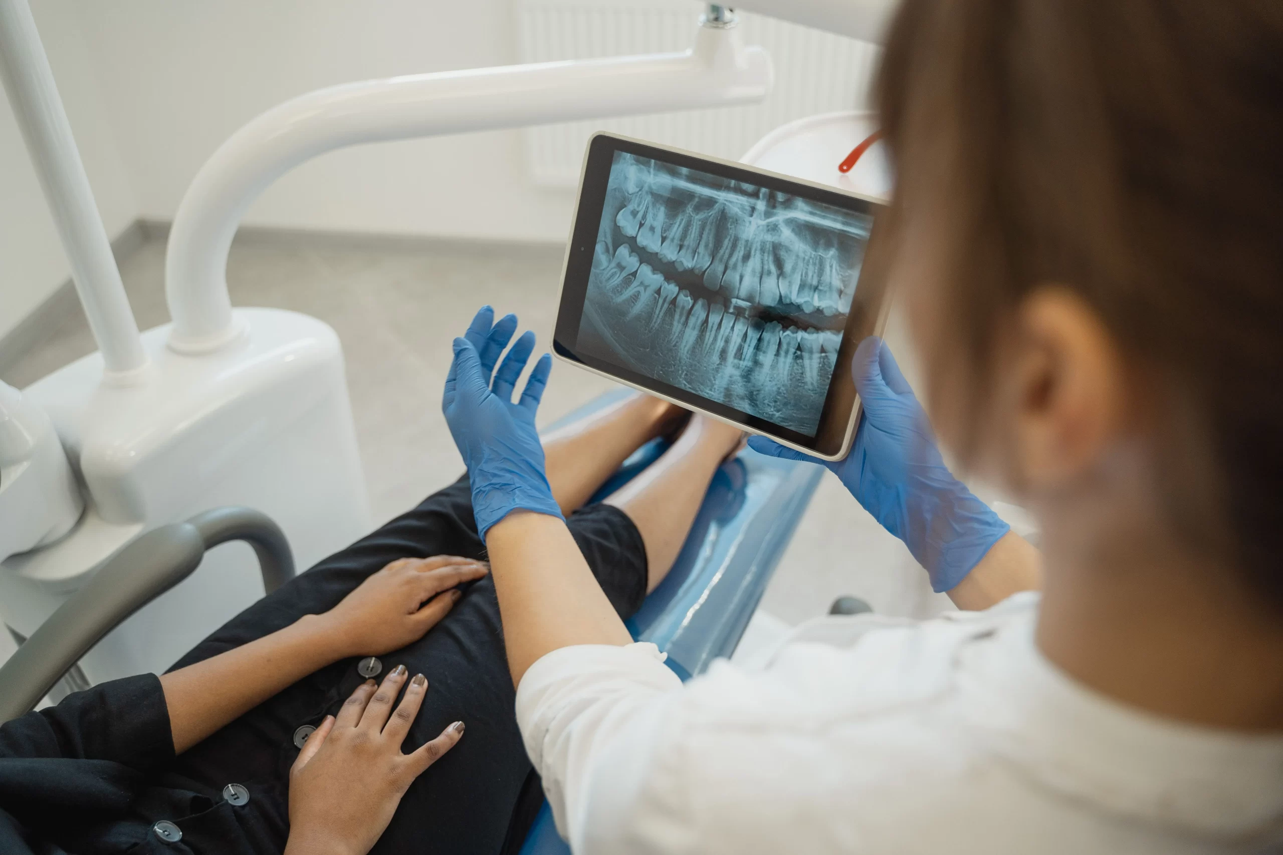
Wisdom teeth are the final adult teeth to develop at the back of the mouth. Some erupt normally and become functional teeth. Others remain partly or completely trapped beneath the gum or within the jawbone.
This is known as wisdom tooth impaction.
Dentists classify impacted wisdom teeth in several ways. They may describe the direction in which the tooth is angled, how deeply it sits in the jaw and whether it has partially erupted through the gum.
The classification provides a useful description of the tooth, not a treatment decision. Symptoms, surrounding anatomy, dental X-rays and the condition of the neighbouring teeth must also be considered.
How wisdom tooth impactions are classified
The position of an impacted wisdom tooth is often described by comparing its angle with the second molar immediately in front of it.
| Type of impaction | Direction of the wisdom tooth | Typical appearance |
| Mesioangular impaction | Crown tilts forwards towards the second molar | Tooth leans into the back of the neighbouring tooth |
| Vertical impaction | Tooth remains relatively upright | Long axis is broadly parallel with the second molar |
| Horizontal impaction | Tooth lies almost sideways | Crown points towards the second molar |
| Distoangular impaction | Crown tilts backwards | Tooth angles away from the second molar |
| Other positions | Tooth angles towards the cheek, tongue or another direction | Position falls outside the four common classifications |
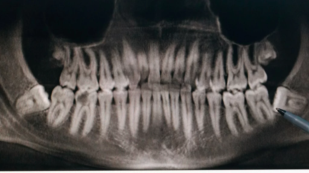
These descriptions are based on established radiographic classification methods. A clinical and radiological study of impacted wisdom teeth explains that angulation, eruption status, depth and surrounding anatomy may all form part of the assessment.
Mesioangular impaction
A mesioangular wisdom tooth tilts forwards towards the second molar. This may also be referred to more generally as a mesial impaction.
Because the crown points towards the tooth in front, plaque and food can sometimes collect between the two teeth. Whether this causes a problem depends on how much of the tooth has erupted, whether the area can be cleaned and whether the neighbouring molar has been affected.
A mesioangular tooth may remain completely beneath the gum or emerge only partly. Its angle alone does not mean that removal is necessary.
Vertical impaction
With a vertical impaction, the wisdom tooth remains relatively upright. It may appear to be following a normal eruption path but still fail to emerge fully because there is not enough space at the back of the jaw.
Some vertically impacted teeth remain symptom-free. Others partially erupt and leave a flap of gum over part of the crown, creating an area that can be difficult to clean.
A vertical position should not be confused with successful eruption. The dentist still needs to assess whether there is enough space, whether the tooth can be maintained and whether the surrounding tissues are healthy.
Horizontal impaction
A horizontal impacted wisdom tooth lies almost sideways within the jaw. Its crown points towards the back surface or roots of the second molar rather than towards the biting surface.
This can place the wisdom tooth close to the neighbouring molar. A dental X-ray may help show whether there are signs of decay, loss of root structure, bone changes or another condition requiring attention.
A horizontal position can make removal more involved in some cases, but angulation is only one consideration. Root shape, bone coverage, depth and proximity to important structures also influence treatment planning.
Distoangular impaction
A distoangular wisdom tooth tilts backwards, away from the second molar.
Depending on its position, it may remain fully within the jaw or partly emerge behind the gum. Cleaning access can be limited because the tooth sits at the very back of the mouth.
Whether it requires attention depends on symptoms, eruption status and surrounding anatomy rather than the angle alone.
Less common positions
Some wisdom teeth develop at angles that fall outside the four common classifications. They may lean towards the cheek or tongue, or develop in an inverted or otherwise unusual position.
The dentist will interpret these positions alongside the tooth’s depth, root shape and relationship with nearby teeth, nerves, bone and other structures.
Partially and fully impacted wisdom teeth
The angle of the tooth is only one part of its classification. Dentists also consider how much of the tooth has erupted.
Partially impacted wisdom tooth
A partially impacted wisdom tooth has started to emerge, but part of its crown remains covered by gum.
This can leave a small pocket beneath the gum flap where plaque and food debris collect. The tissues may become inflamed, particularly when the area is difficult to reach with a toothbrush.
Possible symptoms include:
- tenderness behind the second molar
- red or swollen gum
- an unpleasant taste
- discomfort when chewing
- difficulty opening the mouth fully
- repeated inflammation around the tooth
Discomfort from an inflamed wisdom tooth may be felt in nearby parts of the jaw or face. Symptoms with another possible cause still require appropriate assessment.
Fully impacted wisdom tooth
A fully impacted wisdom tooth has not broken through the gum. It may be covered by soft tissue, jawbone or both.
Because it is not visible in the mouth, you may be unaware of the tooth until it appears on an X-ray.
A fully impacted tooth is not automatically unhealthy and does not necessarily require removal. Monitoring may be appropriate when the tooth is not causing symptoms, and there is no evidence of disease.
Regular dental check-ups allow the tooth and neighbouring molar to be reviewed over time.
Soft-tissue and bony impactions
Impacted wisdom teeth may also be classified by the type of tissue covering the crown.
- Soft-tissue impaction: The tooth has moved beyond the bone but remains partly or fully covered by gum.
- Partial bony impaction: Part of the crown remains surrounded by jawbone.
- Complete bony impaction: The tooth is fully enclosed within bone.
These descriptions can help a dentist or oral surgeon understand how accessible the tooth may be. They do not provide a complete prediction of surgical difficulty or risk.
What can a dental X-ray show?
A clinical examination may reveal whether the tooth is visible, partially erupted or surrounded by inflamed gum. Imaging provides information about structures that cannot be seen directly.
Healthdirect’s guidance on removing wisdom teeth explains that wisdom teeth may partially erupt or become impacted against another tooth or surrounding bone.
Depending on the type and quality of the image, a dental X-ray may help the dentist assess:
- the direction in which the tooth is angled
- how deeply it sits in the jaw
- whether it contacts the second molar
- the number and shape of its roots
- the amount of surrounding bone
- signs of decay, infection, root damage or cystic change
- the apparent relationship between a lower wisdom tooth and nearby nerves
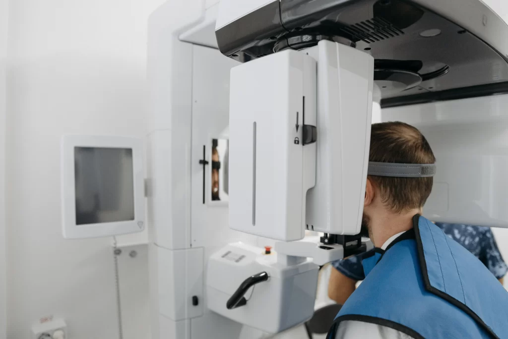
In selected cases, further imaging may be considered when a standard dental X-ray does not answer a specific question needed for treatment planning. Additional imaging is not required for every impacted wisdom tooth.
Does every impacted wisdom tooth need to be removed?
No. An impacted wisdom tooth does not necessarily need to be removed simply because it is angled, buried or unable to erupt fully.
The Better Health Channel’s guidance on wisdom teeth notes that some wisdom teeth do not cause problems and do not need to be taken out.
A dentist will consider whether the tooth is causing symptoms, is difficult to maintain or shows signs of disease before recommending treatment.
Clinical reasons that may lead a dentist or oral surgeon to discuss removal include:
- repeated or serious infection around the tooth
- decay that cannot be predictably restored
- decay or damage affecting the second molar
- an abscess
- certain cysts or other pathological changes
- damage or loss of root structure affecting the wisdom tooth or neighbouring tooth, known as root resorption
- obstruction of another necessary dental procedure
One episode of inflammation does not establish by itself that removal is necessary. Its severity, recurrence, cleaning access and the clinical and X-ray findings all need to be considered.
Monitoring may be recommended when the tooth is symptom-free, and no disease is identified.
How is the treatment approach decided?
Angulation is only one part of treatment planning.
The dentist may also consider:
- the nature and frequency of your symptoms
- whether the tooth and surrounding area can be cleaned
- the condition of the second molar
- the shape and development of the roots
- the amount of bone covering the tooth
- the tooth’s relationship with nearby nerves or the sinus
- relevant health conditions and medicines
- the expected risks and benefits of monitoring or removal
When removal is appropriate, the procedure may sometimes be carried out using local anaesthetic. Sedation or another form of anaesthesia may be considered according to the complexity of the procedure, your medical suitability and your level of anxiety.
Our guide to dental sedation options explains the general approaches that may be discussed.
No sedation method is suitable for everyone, and an individual assessment is required. Sedation also does not remove the need for local anaesthetic where this is clinically necessary.
When should an impacted wisdom tooth be checked?
Arrange a dental assessment if you notice persistent or recurring symptoms around the back of the mouth, such as:
- pain or tenderness
- repeated gum swelling
- food becoming trapped around the tooth
- an unpleasant taste or discharge
- difficulty opening your mouth
- discomfort when biting
- visible decay in the wisdom tooth or neighbouring molar
Seek urgent dental or medical advice if swelling is spreading, you feel generally unwell, or swallowing becomes difficult.
If your breathing is affected, call Triple Zero (000) immediately.
How the findings guide monitoring or treatment
The different types of wisdom teeth impactions describe whether a tooth is mesioangular, vertical, horizontal, distoangular or positioned in another direction. A tooth may also be partially erupted, fully impacted or covered by different amounts of gum and bone.
These classifications help describe the tooth, but they do not determine treatment by themselves. Symptoms, examination findings, imaging, cleaning access and the presence or absence of disease all need to be considered.
If you are concerned about an impacted or partially erupted tooth, visit our wisdom tooth removal service to learn more about assessment and possible treatment options.
An examination and suitable imaging can help determine whether monitoring, treatment or referral is appropriate for your circumstances.
This article provides general information only and does not replace a dental examination, diagnosis or personalised advice from a registered dental practitioner. Whether an impacted wisdom tooth should be monitored or treated depends on your symptoms, clinical findings, imaging, surrounding anatomy and wider health. All surgical and invasive procedures carry risks, which should be discussed before treatment.