Signs of Enamel Erosion: What to Look For and Why It Matters
Enamel erosion is one of the most frequently observed structural changes during routine dental examinations, often detected before symptoms become apparent. Despite this, it commonly goes unnoticed until sensitivity or visible changes become difficult to ignore.
Enamel is the hard protective surface of the tooth, and once it is lost, the body cannot replace it. Recognising the early signs of enamel erosion matters because it allows protective steps to be considered before enamel loss begins affecting tooth strength or appearance.
What enamel erosion actually is
Tooth enamel is the outermost, mineralised layer that shields each tooth from daily wear and chemical attack. Enamel erosion occurs when acids repeatedly soften and dissolve this protective layer over time. Unlike tooth decay, which is driven by bacterial activity, enamel erosion is caused directly by acid exposure.
As enamel thins, the dentine beneath becomes exposed. Dentine is naturally darker in colour and contains microscopic tubules that transmit sensations directly to the tooth nerve. This explains why sensitivity is often one of the earliest and most noticeable symptoms.
Early signs of enamel erosion to watch for
Catching enamel erosion early allows for more conservative care and better long-term outcomes. These are the signs that tend to matter most clinically when assessing possible enamel erosion.
Sensitivity to temperature, sweet or acidic foods
A sharp or sudden sensation when teeth are exposed to cold drinks, hot foods, sugary items, or acidic foods is often an early indicator. This occurs because exposed dentine transmits stimuli more readily than enamel. Medical guidance on tooth sensitivity and exposed dentine explains how enamel loss increases nerve response to thermal and chemical triggers.
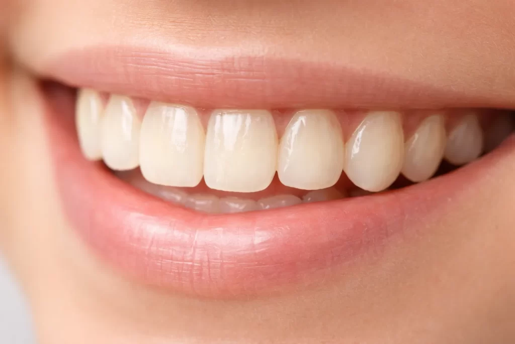
Teeth appear more yellow
As enamel becomes thinner, the underlying dentine shows through more clearly. This change is structural rather than surface staining and is frequently mistaken for discolouration, even when oral hygiene habits have not changed.
Smooth or glossy tooth surfaces
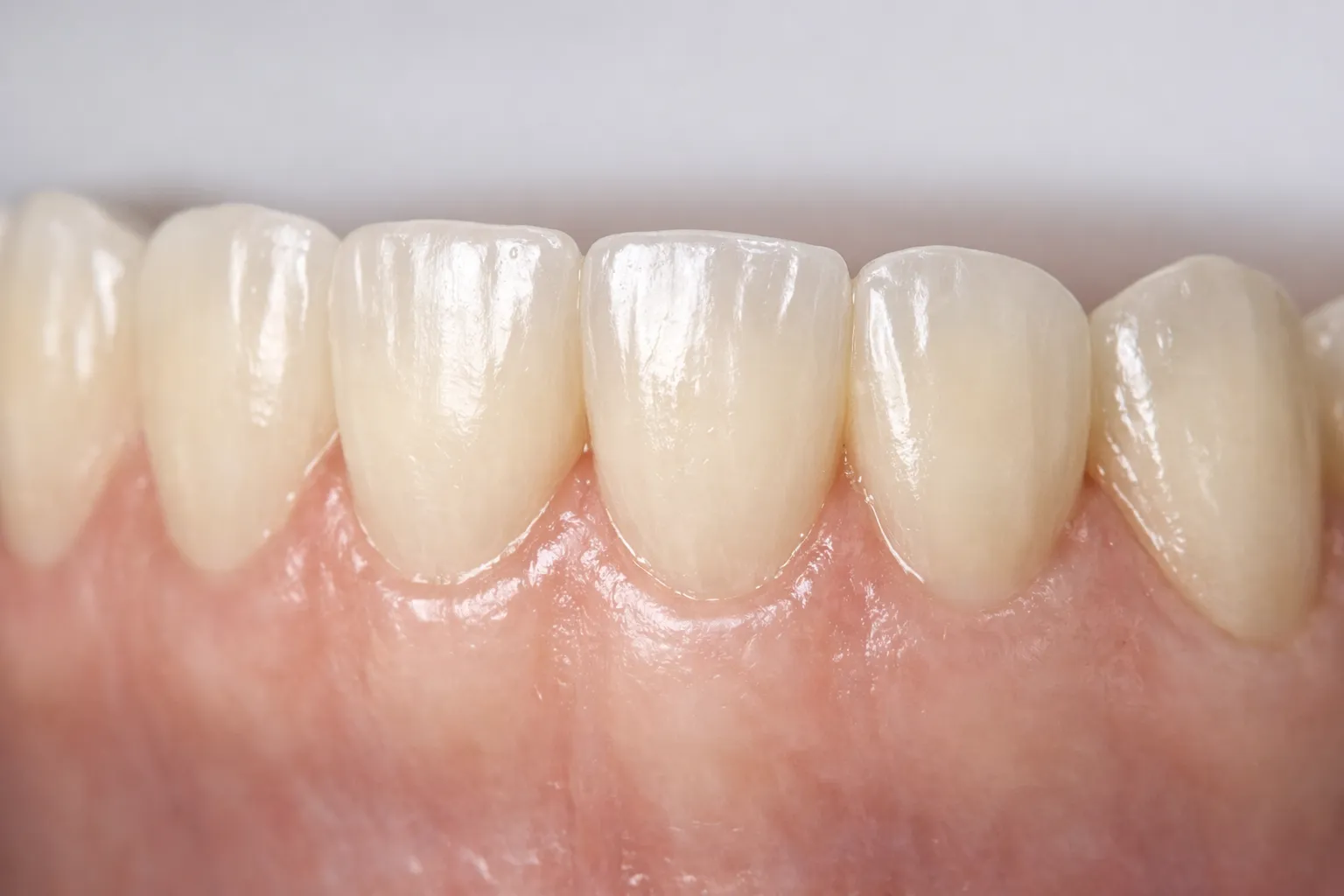
Healthy enamel has a subtle surface texture. With erosion, teeth may develop an unusually smooth or polished appearance, particularly on the front teeth and chewing surfaces. This smoothing and loss of surface detail is a recognised feature of erosive tooth wear described in clinical reviews of erosive tooth wear characteristics.
Transparent edges on front teeth
The biting edges of the front teeth may begin to look translucent or glass-like. This is one of the clearer visual signs of thinning enamel and is often easiest to notice in bright lighting.
Rounded or softened tooth shape
As erosion progresses, natural tooth contours can lose definition. Edges that were once crisp may appear rounded, and biting surfaces can look flatter than they once did.
Cupping on the chewing surfaces
On back teeth, enamel erosion often presents as shallow dents or cupping on the chewing surfaces. These changes are rarely felt by the patient and are commonly identified during a clinical examination rather than through symptoms alone.
Increased chipping or fine cracks
Thinner enamel provides less protection against everyday biting forces. Minor chips or fine cracks may begin appearing more easily, particularly at the edges of the teeth.
Fillings are becoming more noticeable
Because fillings do not erode like natural enamel, surrounding tooth structure may wear away, making restorations feel raised or appear more prominent over time.
Teeth appearing shorter
In more advanced cases, gradual enamel loss can reduce tooth height, particularly in areas under heavier chewing load. This can subtly alter how the bite feels as well as how the teeth look.
Is it enamel erosion or something else?
Not every sensitive or worn tooth is caused by enamel erosion. Distinguishing between erosion, decay, and mechanical wear helps guide the right management approach.
| What you notice | Likely explanation |
| Sensitivity to cold or sweet foods | Erosion, decay, or exposed dentine |
| Yellowing without obvious staining | Enamel erosion |
| Smooth or glossy surfaces | Enamel erosion |
| Localised pain with visible cavities | Tooth decay |
| Flattened biting surfaces | Grinding or abrasion |
Because these conditions can overlap, an in-person assessment is often needed. The distinction between structural wear and decay is explored further in Decay and Missing Teeth: All Your Questions Answered in the Chair.
Common causes of enamel erosion
Enamel erosion usually follows a recognisable pattern: repeated acid exposure without enough recovery time.
Dietary acids
Frequent exposure to acidic foods and drinks is one of the leading contributors. What matters most is not the occasional acidic item, but how often acids contact the teeth. Research examining dietary acids and erosive tooth wear shows that frequent exposure significantly increases enamel loss over time, particularly when acids are consumed throughout the day.
Stomach acid exposure
Stomach acid is significantly stronger than most dietary acids. Reflux can expose teeth to acid from within the body, often affecting the inner surfaces first. This is one reason enamel erosion can progress quietly before it becomes obvious.
Reduced saliva flow
Saliva neutralises acids and supports remineralisation. When saliva flow is reduced due to dehydration, medications, dry mouth, or mouth breathing, acids remain on the tooth surface longer and increase erosion risk.
Brushing habits
Brushing immediately after acidic exposure can worsen enamel loss because the enamel surface is temporarily softened. Excessive pressure or abrasive toothpaste can compound this effect.
What to do if signs of enamel erosion are noticed
Early action can help slow progression and reduce discomfort.
Step 1: Delay brushing after acidic intake
Rinsing with water and waiting around 30 minutes before brushing is commonly recommended to allow saliva to neutralise acids.
Step 2: Reduce acid contact frequency
Avoid sipping acidic drinks over long periods. Having them with meals or finishing with water can help reduce ongoing exposure.
Step 3: Review brushing technique
Using a soft toothbrush, gentle pressure, and a low-abrasive toothpaste is generally advised to minimise further wear.
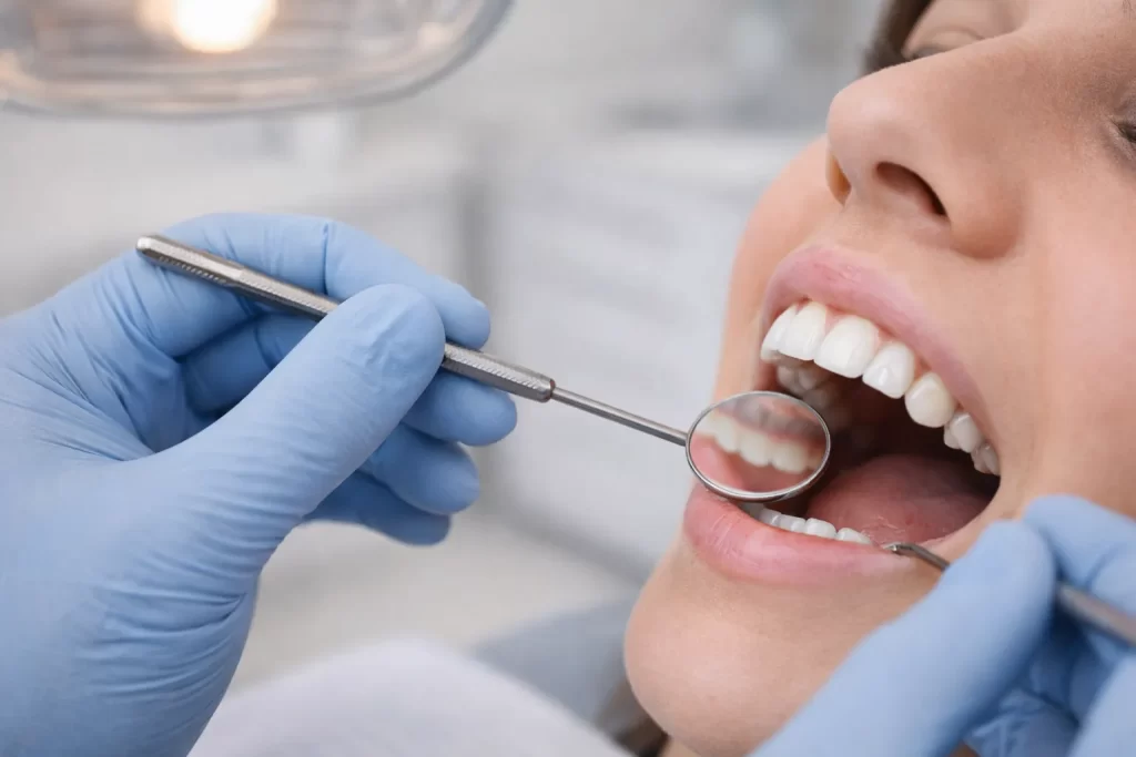
Step 4: Arrange a professional assessment
A proper assessment looks at enamel thickness, surface texture, sensitivity response, bite patterns, and contributing habits to determine whether erosion is active or stable. Guidance on how often you should visit the dentist explains why these subtle changes are best monitored over time.
Where preventive dental care fits in
Enamel erosion cannot be assessed accurately from symptoms alone. Only a clinical examination can determine whether enamel erosion is present and whether it is active or stable.
This type of evaluation is a core part of general dental care, where the emphasis is on early identification and protecting tooth structure before erosion progresses into more invasive treatment.
If sensitivity, translucency along the edges of the teeth, or changes in tooth shape are being noticed, an in-person assessment becomes important. Contact the practice to arrange a check-up and discuss how early intervention may help preserve enamel.
Key takeaway
Enamel erosion develops gradually and often without obvious early symptoms. Sensitivity, colour changes, surface smoothing, and altered tooth shape are important warning signs. While enamel cannot be replaced, early identification and targeted care may help slow progression and reduce the likelihood of more complex treatment later in some cases.
This information is general in nature and does not replace an individual dental assessment.